
Muscle Condition Score (MCS) for Dogs and Cats: A Clinical Guide
Published and maintained by the World Pet Obesity Association (WPOA)
Muscle Condition Score (MCS) is a visual and palpation-based assessment of muscle mass in dogs and cats. It complements Body Condition Score (BCS), which assesses body fat. Record MCS as normal muscle mass or mild, moderate, or severe muscle loss. MCS is a clinical assessment, not a direct measurement of total lean body mass. A dog or cat can have excess body fat and clinically important muscle loss at the same time.
This World Pet Obesity Association (WPOA) clinical guide presents a five-site workflow for dogs and cats, a four-category descriptive scale, species-specific considerations, documentation guidance, and ways to integrate MCS into weight-management care and client communication. Use MCS alongside body weight and BCS during routine nutritional assessment.
Independent resource notice
This complete guide and chart are WPOA resources. WPOA is responsible for the clinical content, chart, updates, translations, downloads, and related guidance. APOP provides access to this guide as an external veterinary education resource. APOP and WPOA are independently operated organizations.
Clinical Resource
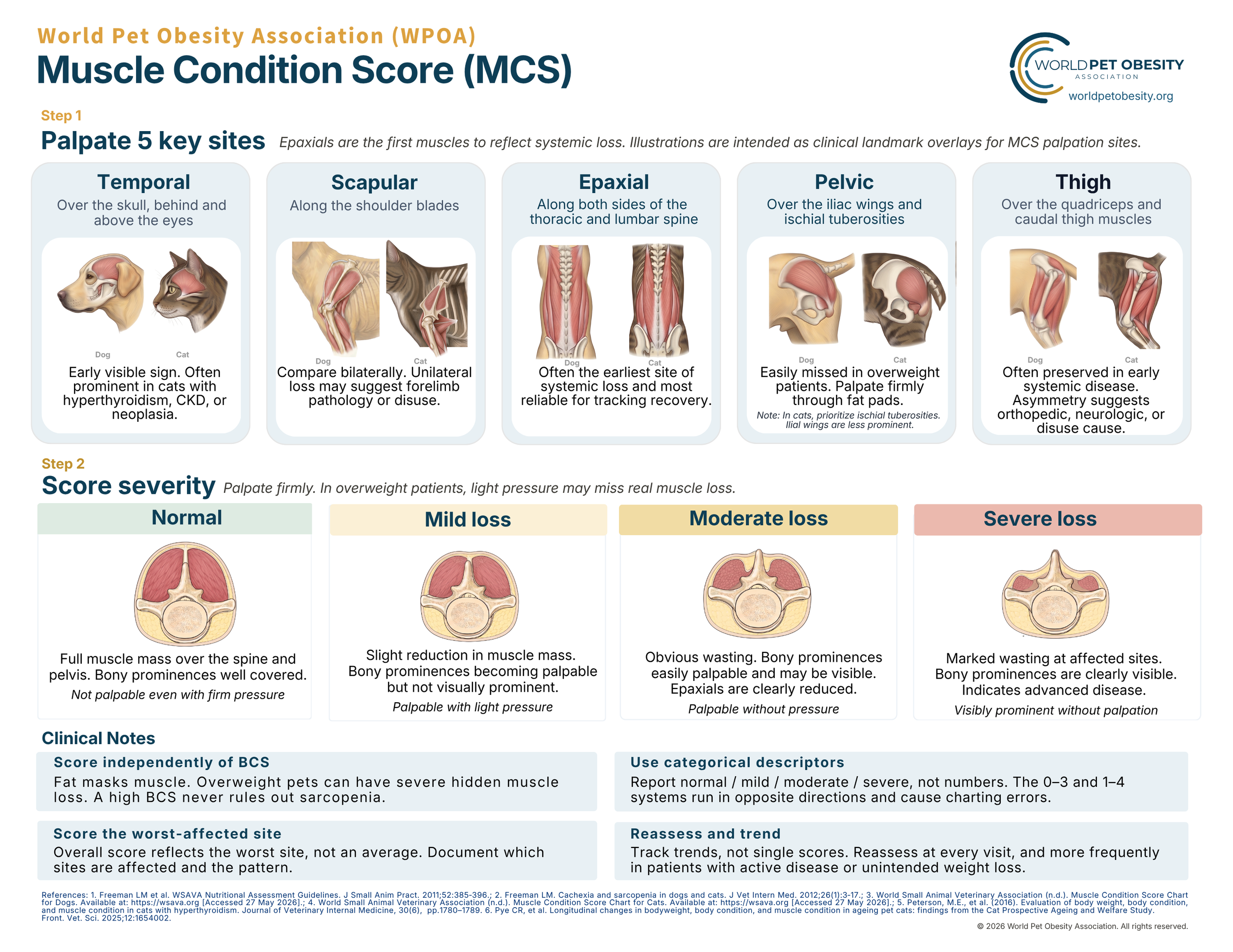
MCS Chart for Dogs and Cats
Clinic-ready PDF with palpation sites, severity descriptors, and documentation guidance. Print for exam rooms or share with your team.
Quick facts: Dogs and cats · 4 descriptive categories · Visual assessment and palpation · WPOA five-site workflow · Use alongside BCS and body weight
Why MCS Matters
BCS does not assess muscle. Patients with a high BCS can present with concurrent moderate-to-severe muscle loss. Without MCS, this finding goes undocumented.
Weight-loss plans can reduce muscle as well as fat. Serial MCS at rechecks helps identify declining muscle condition and supports reassessment of diet, intake, activity, and health.
Muscle loss is clinically important. It is associated with impaired function and poorer outcomes in several chronic diseases, but MCS alone does not establish the cause or prognosis.
Age-related muscle loss can occur in senior dogs and cats. Body weight may remain stable while muscle condition declines, so MCS provides information the scale alone cannot.
MCS supports clearer communication. Describing muscle condition alongside body fat helps clients understand why body weight alone does not show the whole patient.
Scoring Categories
Use the four descriptive categories below to record muscle condition. In the WPOA five-site workflow, palpate all five sites, document every affected site, and assign the overall category from the site with the greatest loss. Do not average categories across sites. This is a clinical documentation method, not a direct measurement of total muscle mass.
| Score | Finding | Palpation |
|---|---|---|
| Normal | Expected muscle volume and contour at the assessed regions. Bony landmarks are appropriately covered for the individual patient. | No clinically apparent muscle loss on visual assessment and palpation |
| Mild loss | Subtle reduction in muscle volume. Bony landmarks may be easier to feel but are not clearly prominent. | Slight loss of expected contour compared with the individual patient or prior examinations |
| Moderate loss | Clear reduction in muscle volume. Bony landmarks are readily palpable and may be visible. Regional concavity or reduced epaxial fullness may be present. | Definite muscle loss at one or more assessed sites |
| Severe loss | Marked reduction at one or more sites with prominent bony landmarks, pronounced concavity, or substantial loss of regional muscle volume. | Severe loss apparent on visual assessment and palpation |
Use the descriptor: normal muscle mass, mild loss, moderate loss, or severe loss. Numeric systems vary among sources. If a number is used, name the scoring system and include the descriptive category to reduce ambiguity.
Palpation Technique
MCS requires both visual assessment and palpation. Coat, body fat, posture, conformation, and positioning can obscure muscle loss. Use enough pressure to assess through coat and subcutaneous fat without causing discomfort. Do not treat a particular pressure level as a validated scoring cutoff.
The five key regions
Temporal
Assess the temporalis region over the skull, behind and above the eyes. Compare both sides and note reduced fullness or concavity.
Scapular
Palpate along and around the shoulder blades. Compare both sides. Focal asymmetry may reflect orthopedic, neurologic, painful, or disuse-related change.
Epaxial
Palpate the muscles along both sides of the thoracic and lumbar spine. The epaxial region is often useful for tracking change over time.
Pelvic
Assess over the wings of the ilia and ischial tuberosities. Excess body fat can obscure loss, so careful palpation is important.
Thigh
Assess quadriceps and caudal thigh muscle volume and symmetry. Asymmetry may suggest orthopedic, neurologic, painful, or disuse-related change.
Rib and intercostal muscle coverage can provide additional context, but these areas are not among the five named sites on the WPOA chart. Interpret them with the complete examination.
Performing Muscle Condition Scoring
- Position Perform on a non-slip surface, with weight even across all four limbs. Sternal recumbency is acceptable if standing isn't possible. Lateral recumbency can be less reliable.
- Inspect from above and the side Note temporal fossa depth, scapular contour, spinous process prominence, and any concavity flanking the spine.
- Palpate the five sites Use the WPOA sequence: Temporal → Scapular → Epaxial → Pelvic → Thigh. Compare both sides and interpret findings against the expected muscle for species, breed, body size, life stage, health, and conditioning.
- Assess ribs and intercostals Evaluate muscle coverage at the ribs, especially intercostal spaces.
- Apply the WPOA documentation method Record all affected sites. In this five-site workflow, assign the overall category from the site with the greatest loss rather than averaging categories across sites.
Dog and Cat Considerations
Dogs
Expected muscle contour varies widely with breed, body size, conformation, age, and conditioning. Long coats and excess body fat can hide muscle loss. Compare both sides, and interpret focal asymmetry in the context of pain, mobility, orthopedic disease, neurologic disease, and disuse.
Cats
The same WPOA five-site workflow can be used in cats, but the examination often requires different emphasis. Cats may lose clinically important muscle while maintaining an ideal, overweight, or obese BCS, so body weight and visual inspection can be misleading. Carefully assess the epaxial and temporal regions and interpret any concavity or reduced fullness with the complete MCS examination.
The ventral fat pad and subcutaneous fat can mask clinically important muscle loss. A cat may have BCS 8-9/9 and still have moderate-to-severe muscle loss. Body Condition Score assesses body fat; Muscle Condition Score assesses muscle condition through visual assessment and palpation.
In some cats, the ischial tuberosities may be easier to assess than the ilial wings.
Cats with chronic disease may lose clinically important muscle while maintaining an ideal, overweight, or obese BCS. Assess all five regions and document the affected sites and severity.
Matted coat over the dorsum, tail base, and hindquarters may reflect reduced flexibility, pain, weakness, or systemic disease. When present with muscle loss, document both findings and consider whether mobility limitation or underlying disease is contributing.
The primordial pouch is not a muscle landmark and should not be confused with body or muscle condition findings.
Documentation
Record MCS alongside body weight and BCS during routine nutritional assessment and at clinically appropriate rechecks. Trends matter as much as a single finding. Serial assessments can show whether muscle condition is stable, improving, or declining.
Include
- BCS scale used, 5- or 9-point, with the value
- MCS descriptor in words: normal, mild loss, moderate loss, severe loss
- Site(s) of muscle loss and the site of greatest loss when present
- Asymmetry, if observed, which may suggest orthopedic, neurologic, or disuse cause
- Weight trend from the previous visit
Avoid
- An unexplained numeric MCS, such as "MCS 3/4." If a numeric system is used, name the system and include the descriptive category
- "Muscle wasting" without a severity descriptor or affected location. That is too vague to track
- MCS without a paired BCS. They describe different tissues and should be interpreted together. A high BCS does not rule out muscle loss.
Clinical Response Guide
MCS in Weight Management
Before starting
- Record baseline body weight, BCS, and MCS.
- If MCS shows loss at baseline with elevated BCS, document concurrent excess adiposity and muscle loss. Prioritize muscle preservation and evaluate possible underlying causes.
- Choose a nutritionally appropriate plan for the individual dog or cat. When substantial calorie restriction is required, consider a therapeutic weight-management diet or another complete plan that maintains essential nutrient intake.
During
- Reassess body weight, BCS, MCS, measured intake, appetite, activity, mobility, and clinical signs at intervals based on clinical risk and the intended rate of change.
- Use species-appropriate, individualized rate targets established in the weight-management plan. Reduced or absent food intake in cats requires prompt attention.
If MCS declines during weight management, reassess the plan. Review diet adequacy, actual intake, the rate of restriction, activity, pain, and underlying health before continuing unchanged.
After goal weight
MCS should be stable or improved when clinically feasible. Persistent or worsening muscle loss warrants reassessment of the patient and the plan. Continue periodic MCS monitoring during maintenance.
Download the Chart
Clinic-ready resource. Print for exam rooms, share with your team, or download as a PNG for digital use.
References
- Freeman LM, et al. WSAVA Nutritional Assessment Guidelines. J Small Anim Pract. 2011;52:385–396.
- Freeman LM. Cachexia and sarcopenia in dogs and cats. J Vet Intern Med. 2012;26(1):3–17.
- World Small Animal Veterinary Association. Muscle Condition Score Chart for Dogs. wsava.org [accessed 27 May 2026].
- World Small Animal Veterinary Association. Muscle Condition Score Chart for Cats. wsava.org [accessed 27 May 2026].
- Peterson ME, et al. Evaluation of body weight, body condition, and muscle condition in cats with hyperthyroidism. J Vet Intern Med. 2016;30(6):1780–1789.
- Pye CR, et al. Longitudinal changes in bodyweight, body condition, and muscle condition in ageing pet cats: findings from the Cat Prospective Ageing and Welfare Study. Front Vet Sci. 2025;12:1654002.